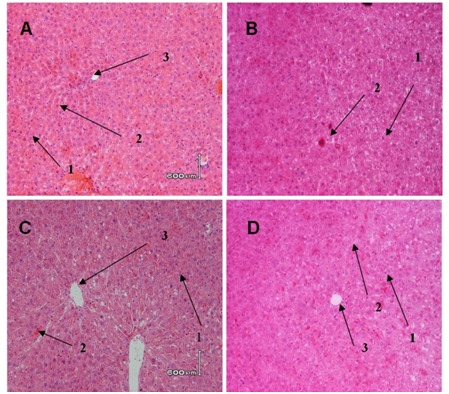
Figure 5.

Histological evaluation in liver tissues of control and experimental group.
Rats were divided into 4 groups, (A) 0.5 % CMC-Na, (B) DMBA control group (20 mg/kgbW in CMC-Na), (C) DMBA+ 300 mg/kgBW EGP; (D) DMBA+750 mg/kgbw EGP. At autopsy, liver organ were removed and fixed in 10% buffered formalin. 3-5 μm tissue slices were embedded in paraffin, and stained with hematoxylin and eosin (a1=hepatocyte, 2=sinusoid,3=central venous). Magnification x 400.
