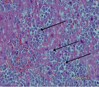
Figure 6.

Hematoxylin and eosin stained of lymphoblastic cells in liver tissues
of DMBA control group (20 mg/kgbW in CMC-Na). At autopsy, liver organ were removed and fixed in 10% buffered formalin. 3-5 μm tissue slices were embedded in paraffin, and stained with hematoxylin and eosin. Lymphoblastic cells are pointed by black arrow. Magnification x 400.
