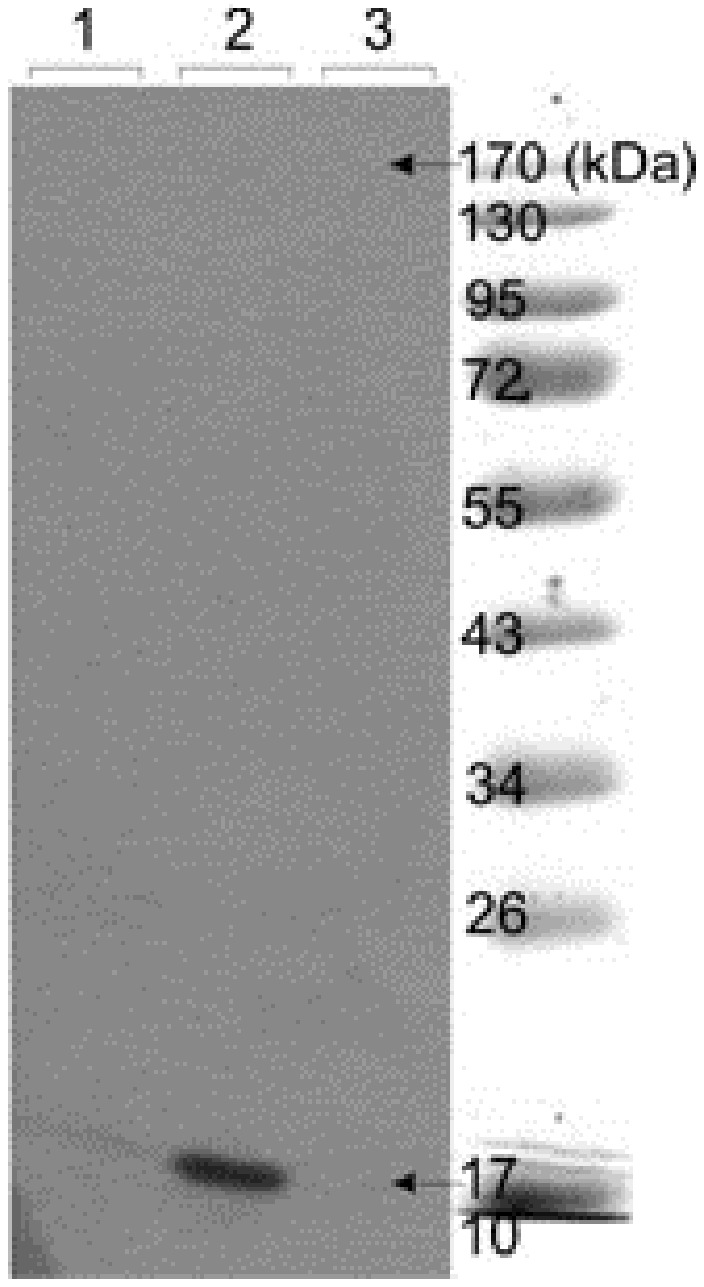
Figure 2.

Western blot analysis of TNF-α in Raji cell lysates. Cultured human Raji cells were stimulated with 10 mg/mL LPS. SDS-PAGE was subjected onto cell lysates and blotted onto PVDF membrane. Lanes 1 and 3: Control. Lane 2: The detected band showing induction of the TNF-α.
