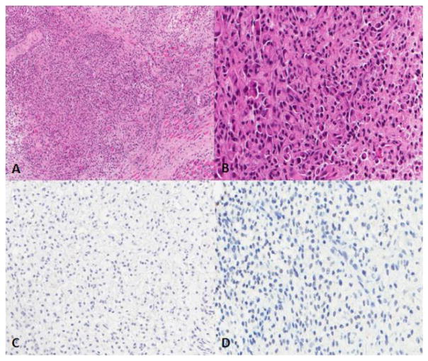
Figure 1.
The tumor consists of poorly differentiated, discohesive, medium-sized polygonal and short spindled cells with nuclear irregularity and atypia. Some of the tumor cells feature eccentric nuclei and abundant eosinophilic cytoplasm. SMARCB1 and Myogenin are negative. A. Hematoxylin and eosin (H&E) stain, 10X; B. H&E stain, 40X; C. Myogenin, 20X; D. SMARCB1, 20X.

