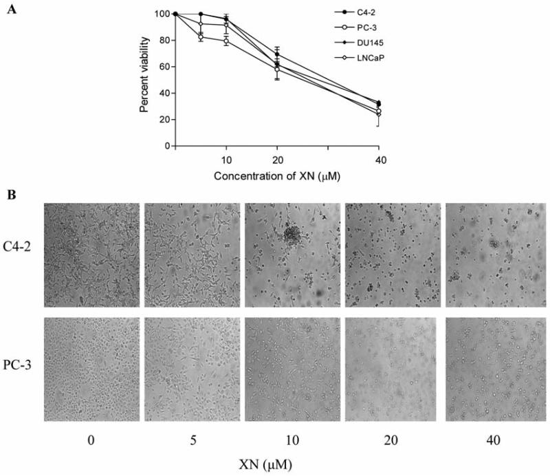
Figure 1.
Effect of XN on the viability of prostate cancer cells. A: 1×10⁴ LNCaP, C4-2, PC-3 and DU145 cells were separately seeded in each well of a microtiter plate in 0.1 ml of culture medium. Cells were allowed to adhere for 24 h before treating with XN at concentrations of 0 to 40 μM in triplicates for 72 h. Cell viability was measured by MTS assay using CellTiter AQueous assay system from Promega. Similar results were obtained in 3 independent experiments. B: Morphological changes in cell cultures (C4-2 and PC-3 cells) treated or not with XN for 72 h as visualized by light microscopy. Similar results were obtained in two independent experiments.

