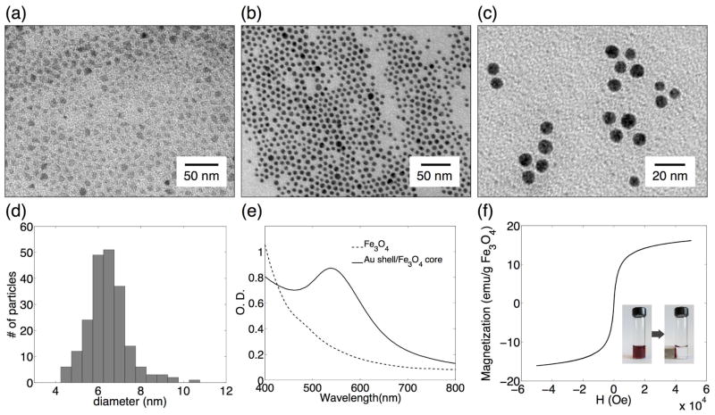
Figure 2.
Characterization of magnetic core/shell nanocarriers. TEM images of Fe3O4 nanoparticles in hexane before (a) and after (b) coating with gold shell; gold shell/magnetic core nanoparticles after transfer into aqueous phase (c). Gold shell/Fe3O4core nanoparticle size distribution (6.2 ± 0.8 nm) as determined from TEM image analysis of more than 200 particles (d). UV-V is spectrum of oleic acid and oleylamine stabilized Fe3O4 nanoparticles (dashed) and gold shell/magnetic core particles in hexane (solid) (e). Magnetization hysteresis at 300 K of gold shell/magnetic core nanoparticles (f); the insert: separation of nanoparticles from a colloidal suspension using a magnetic field gradient created by a simple permanent magnet.

