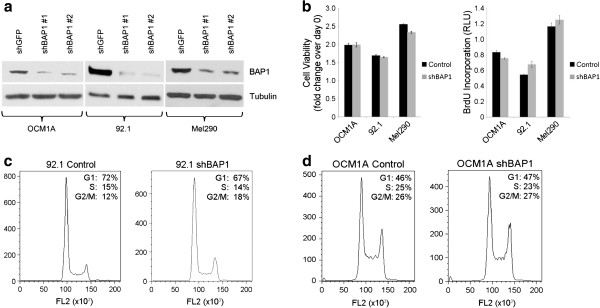
Figure 2.

Stable loss of BAP1 does not promote cell proliferation in uveal melanoma cells. (a) Western blot showing the levels of BAP1 after stable knockdown of BAP1 with two independent lentiviral shRNA constructs in three uveal melanoma cell lines, OCM1A, 92.1, and Mel290. α-tubulin was used as a loading control (b) Cell viability and proliferation of the indicated BAP1-deficient or control stable cells (Left panel) cell viability measured by MTS assay and shown as fold change over day 0 for each stable cell line (Right panel) cell proliferation measured by BrdU incorporation after 24 hrs (c-d) Cell cycle analysis of the indicated cell lines using flow cytometry on propidium iodide stained cells; x-axis represents DNA content and y-axis represents cell number (c) 92.1 stable cells (d) OCM1A stable cells.
