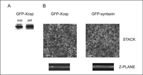
FIGURE 3. Distribution of GFP-csp and GFP-syntaxin 1a in oocytes.
A, an oocyte expressing GFP-csp was fractionated as in Fig. 2, and fractions were analyzed by immunoblot for GFP-csp, which migrates at ~50 kDa. B, image stacks of z-plane sections (obtained by confocal fluorescence microscopy) through cortical region fragments from GFP-csp or GFP-syntaxin 1a expressing oocytes reveal fluorescence surrounding the cortical granules. Representative z-plane sections (side views) show undulating fluorescence associated with the pole of cortical granules closest to the plasma membrane (but not encircling the granules). Scale bar is 5 μm. sup, supernatant; pel, pellet.

