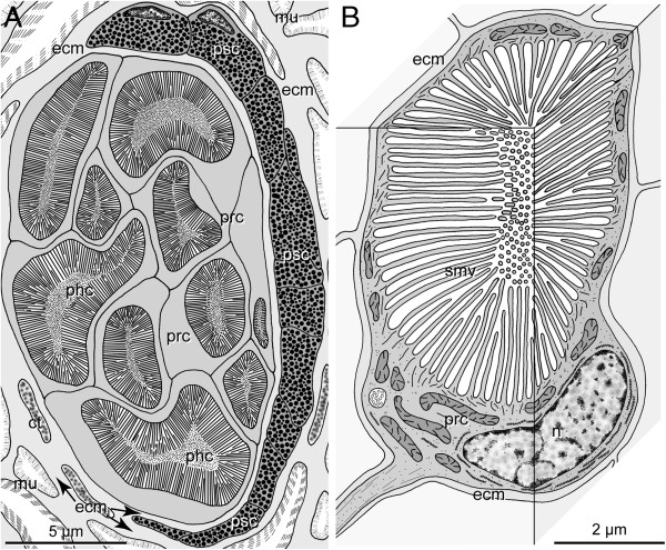
Figure 2.
H. robusta TX; schematic representation of TEM observations of pigmented eyes and PRCs. A. Section through an eye at the level of the phaosomes. The eyes are embedded in the connective tissue (ct) which primarily comprises muscle fibres (mu). Ten PRCs (prc) with their phaosomal cavities (phc) are visible, pigmented supportive cells (psc) form a thin sheath around the PRCs; the two cell types are separated by an ECM (ecm, arrows). B. 3-D view of an extraocular PRC (prc) with its phaosome and sensory microvilli (smv), PRC completely ensheathed by ECM (ecm). - ecm ECM, mu muscle fiber, n nucleus, phc phaosomal cavity, prc photoreceptor cell, psc pigmented supportive cell, smv sensory microvilli.

