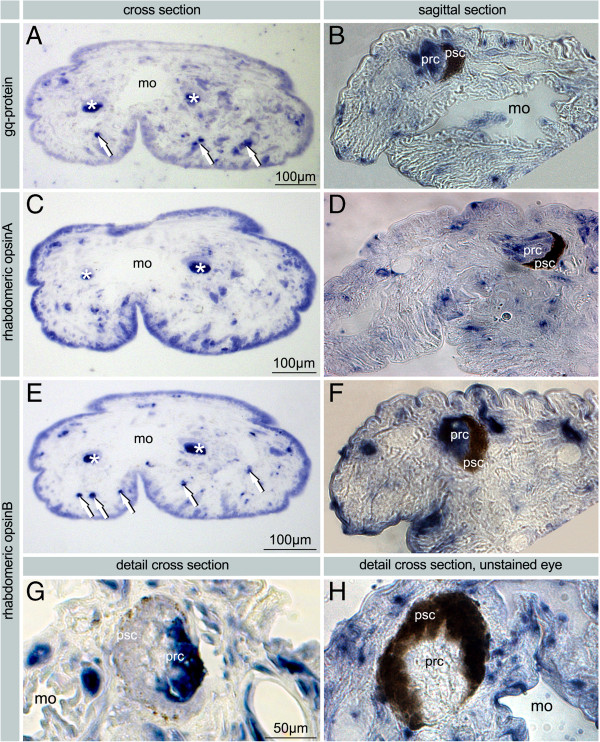
Figure 6.
In situ on paraffin sections of H. robusta using NBT/BCIP. Bright field images. A. Expression of gq-protein (CA) in the PRCs of the eyes (asterisks) and in the extraocular PRCs at the margin of the head (arrows). B. Expression of gq-protein (CA) in the PRCs of the eyes (prc) at higher magnification. C-D. Expression of opsin A (TX) in the PRCs of the eyes (asterisks, prc); and probably in the extraocular PRCs. E-G. Expression of opsin B (TX) in PRCs of the eyes (asterisks, prc) and in the extraocular PRCs (arrows). F. Expression signal in the eyes is restricted to the PRCs G. High magnification image with no expression in PSCs (psc) which lost pigment granules almost entirely during in situ hybridization. Staining of probe is restricted to cell bodies of PRCs, phaosomal cavities remain unstained. H. Sagittal section of unstained specimen showing pigmented eye with pigment granules and no staining in the PRCs. – arrows: extraocular phaosomal PRC; asterisks: pigmented eyes; mo mouth opening; phc phaosomal cavity; psc pigmented supporting cell; px pharynx; smv sensory microvilli.

