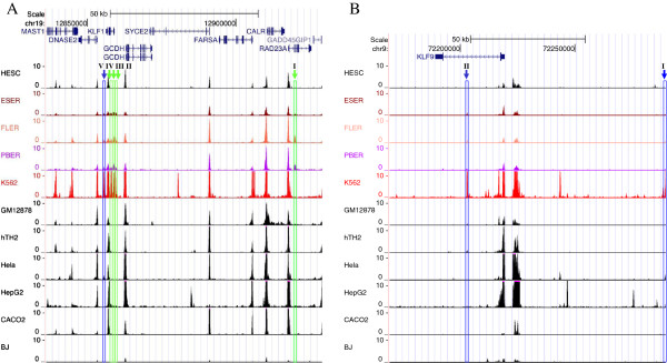
Figure 2.

Distribution of DHSs in the genomic regions of KLF1 and KLF9 genes. Chromatin profiles for KLF1(A) and KLF9(B) are shown to illustrate the distribution of DHSs in the genomic regions of KLF genes in four erythroid (in color) and seven non-erythroid (in black) cell lines. Erythroid-specific or putative erythroid-specific DHSs were named with Roman numbers. Erythroid-specific DHSs, which were present only in human erythroid cells, were indicated by arrows and columns in green. Putative erythroid-specific DHSs, which were also present in non-erythroid cell types but at lower intensities, were indicated by arrows and columns in blue. Therefore, KLF1-I, II, III, and IV (A) were considered as erythroid-specific DHSs, of which KLF1-I was located in the intron region of the RAD23A gene within the defined genomic region, and KLF1-V was a putative erythroid-specific DHS because of its presence in HeLa cells. Similarly, upstream KLF9-I and intronic KLF9-II were considered as putative erythroid-specific sites (B).
