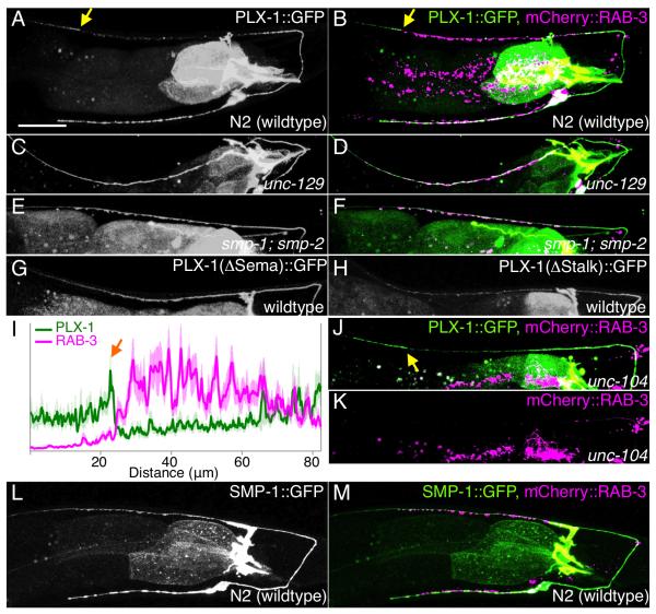
Figure 5. PLX-1::GFP is enriched at a synapse-free domain adjacent to the synaptic region.
PLX-1::GFP localization in wildtype (A, B), in unc-129 mutants with DA9 axon misguided (C, D) and in smp-1;smp-2 mutants (E, F). Each strain has a synaptic marker (Pmig-13::mCherry::rab-3) and merged images are shown in the right panel (B, D and F). (G) PLX-1(ΔSema)::GFP localization in wildtype. (H) PLX-1(ΔStalk)::GFP localization in wildtype. (I) Quantification of the normalized mCherry::RAB-3 signal (magenta) and PLX-1::GFP signal (green) in the dorsal axon of wildtype worms. 18 animals were aligned according to the PLX-1::GFP patch at the anterior edge of the DA9 synaptic domain (orange arrow). Light colors indicate standard error of mean. (J) PLX-1::GFP merged with mCherry::RAB-3 in unc-104/kinesin mutants. mCherry::RAB-3 signal is completely absent from the dorsal axon (K). PLX-1 at the putative tiling border is indicated by yellow arrows. (L) SMP-1::GFP localization in wildtype. (M) mCherry::RAB-3 co-labeled with SMP-1::GFP. Scale bar, 20μm.

