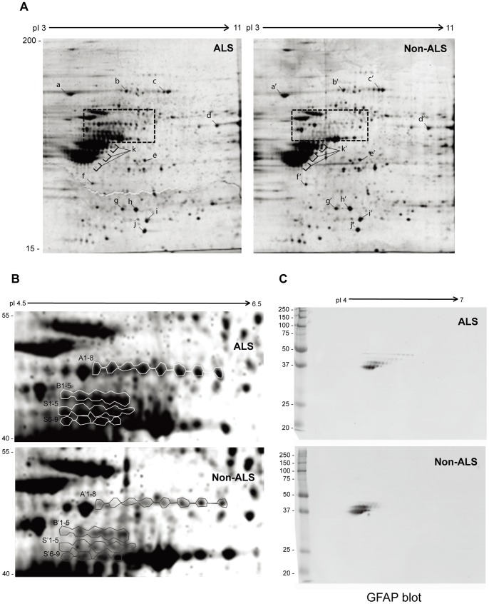
Figure 1. Comparison of urea-soluble proteins from ALS and non-ALS spinal cords by 2D SDS-PAGE.
(A) Urea-soluble whole tissue lysates were prepared from pooled ALS or non-ALS spinal cords in the RIPA lysis buffer containing 8 M of urea. The first dimension was 18-cm immobilized pH gradient isoelectric focusing (IEF) from pI = 3–11; the second dimension was 10% SDS-PAGE. The gels were stained with Sypro Rubby. High quality spots marked with a–i and a’–i’ were randomly selected as references to normalize the differences between different gels. (B) Differentially expressed protein clusters between ALS and non-ALS spinal cords. The cluster A, B and S from ALS and A’, B’ and S’ from non-ALS were excised from the 2-D gels and subjected to LC-MS/MS protein identification. (C) Western blotting analysis of the protein clusters with anti-GFAP antibody. The urea-soluble whole tissue lysates were resolved by mini-2D SDS-PAGE, transferred to the PVDF membrane and detected with the antibody against GFAP.

