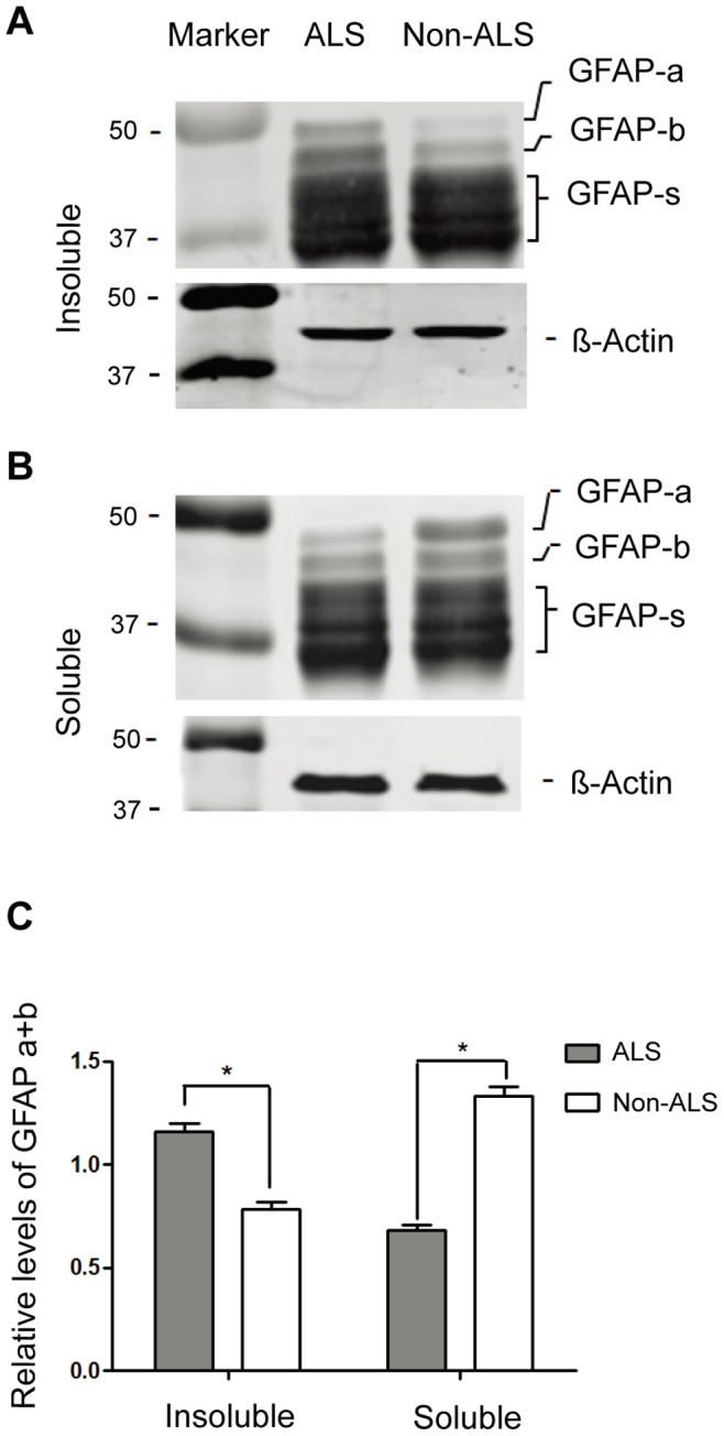
Figure 4. Western Blotting analysis of the GFAP fragments in the soluble and insoluble protein fractions of ALS and non-ALS spinal cords.

(A, B) Western Blotting analysis of the GFAP fragments in the insoluble (A) and soluble (B) fractions. The urea-soluble proteins were dialyzed against PBS and centrifuged. The pellet (insoluble fraction) and the supernatant (soluble fraction) were analyzed by Western blotting and detected with the anti-GFAP antibody. GFAP-a and GFAP-b, the larger forms of GFAP; GFAP-s, the degraded GFAP fragments; β-actin, internal control. (C) Quantitation of two larger forms of GFAP fragments. The expression levels of GFAP-a and GFAP-b relative to β-actin were calculated. The large forms of GFAP are preferably found in the insoluble fractions (* p<0.05, n = 4).
