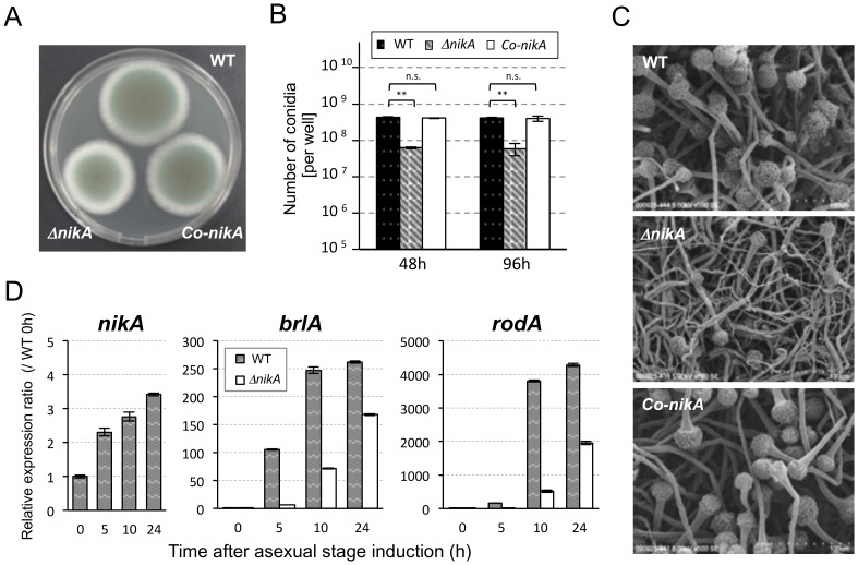
Figure 1. Deletion of the nikA gene results in a conidiation defect.
(A) Conidial suspensions of the parental strain, ΔnikA, and the nikA-complemented strain (Co-nikA) were point-inoculated on 0.1% yeast extract containing glucose minimal medium (YGMM) agar plates and incubated for 68 h at 37°C. (B) The number of conidia in a 34 mm diameter well of a 6-well plate after 48 h and 96 h of incubation at 37°C was counted (See Materials and Methods). Error bars represent the standard deviations based on three independent replicates. **P<0.001 versus WT strain at any time point. P-values were calculated by the Student’s t-test. (n.s.: not significant) (C) The colony surface was observed by scanning electron microscopy (SEM). The growth conditions were the same as those in (B): 48 h incubation at 37°C in 6-well plates. (D) The expression of nikA, brlA, and rodA during the asexual development stage was determined by real-time reverse transcriptase polymerase chain reaction (RT-PCR). To synchronize asexual development initiation, mycelia that were cultured in liquid YGMM for 18 h were harvested and transferred onto YGMM plates (the time point was set as 0 h of the asexual stage). Relative expression ratios were calculated relative to WT at 0 h. Error bars represent the standard deviations based on three independent replicates.

