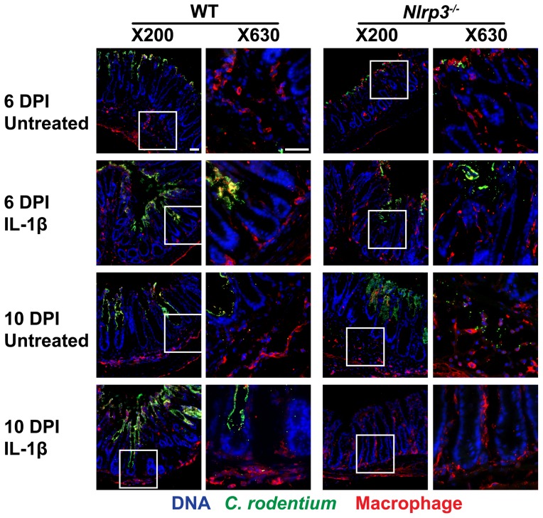
Figure 5. IL-1β treatments augment macrophage colonic infiltration.
Macrophage infiltration was assessed by staining cryosections of distal colons. An increase in the number of macrophages (red; indicated by arrowhead in higher magnification panels on the left) in close proximity of C. rodentium (green) at 10 DPI in Nlrp3−/− mice is noted. With IL-1β treatments, the macrophages in crypts and mucosal lining appeared increased and bacterial infiltration into the crypts reduced. Bar 50 µm.

