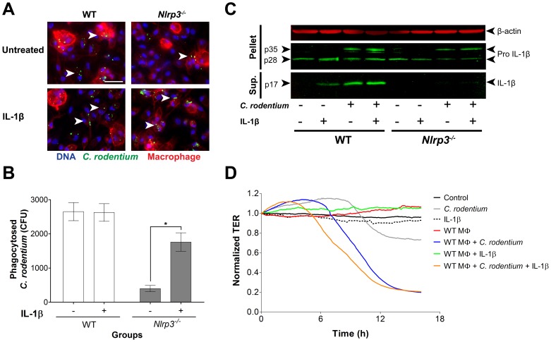
Figure 6. IL-1β enhances C. rodentium phagocytosis, while overcompensation of IL-1β augments epithelial barrier damage in vitro.
(A) Exogenous IL-1β induced C. rodentium phagocytosis (arrowhead) by peritoneal macrophages in vitro, especially in Nlrp3−/− macrophages. Magnification X630, bar 50 µm; (B) Quantification showed an increase of intracellular C. rodentium in IL-1β treated Nlrp3−/− macrophages but no effect on WT macrophages. Data represents mean ± SE. One asterisk P<0.05; (C) Western blot analysis of mature IL-1β in the supernatant of peritoneal macrophages and pro-IL-1β in the cellular component; (D) The epithelial integrity of C. rodentium-infected CMT-93 cells assessed by ECIS, deteriorated in presence WT macrophages and even more with the addition of IL-1β. Data represents the mean of two independent experiments.

