
Figure 1.
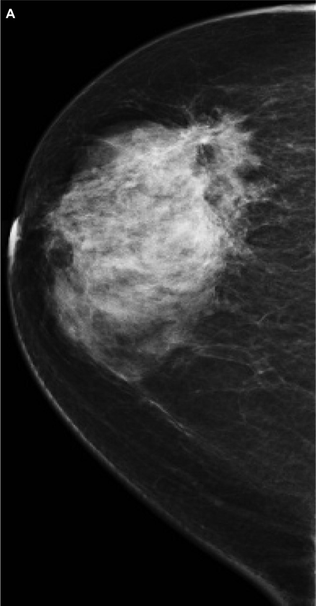
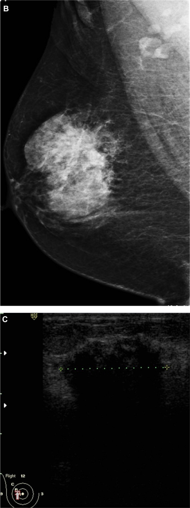
A 51-year-old female presented with hard right breast after suffering from right breast trauma 2 months previously. (A and B) Craniocaudal and mediolateral mammogram of the right breast revealed a dense breast with architectural distortion noted at upper outer quadrant (more evident on craniocaudal view). (C) Ultrasound revealed an ill-defined irregular hypoechoic mass with posterior acoustic shadowing at 9 o’clock of the right breast.
Note: The longitudinal axis of the mass was parallel to that of the skin.