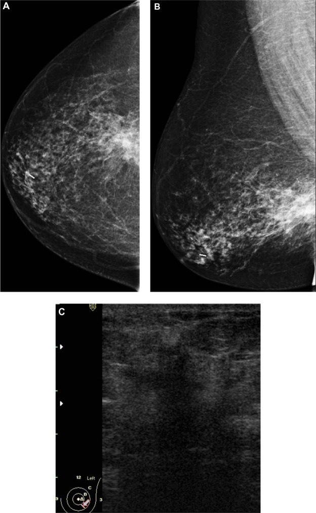
Figure 3.
A 58-year-old female with palpable mass in the left breast. (A and B) CC and ML mammogram films revealed ill-defined dense opacity with spiculated margins surrounded by architectural distortion at the lower central area of the breast, which looks to be attached to the pectoralis muscle posteriorly. (C) On ultrasound, ill-defined, spiculated, hypoechoic, solid mass with posterior shadowing measuring 16 × 13 mm is seen at 5 o’clock of the breast.
Notes: The mass was inseparable from the underlying muscle. Another smaller, similar hypoechoic mass is seen close by measuring 6 × 5 mm.
Abbreviations: CC, craniocaudal; ML, mediolateral.

