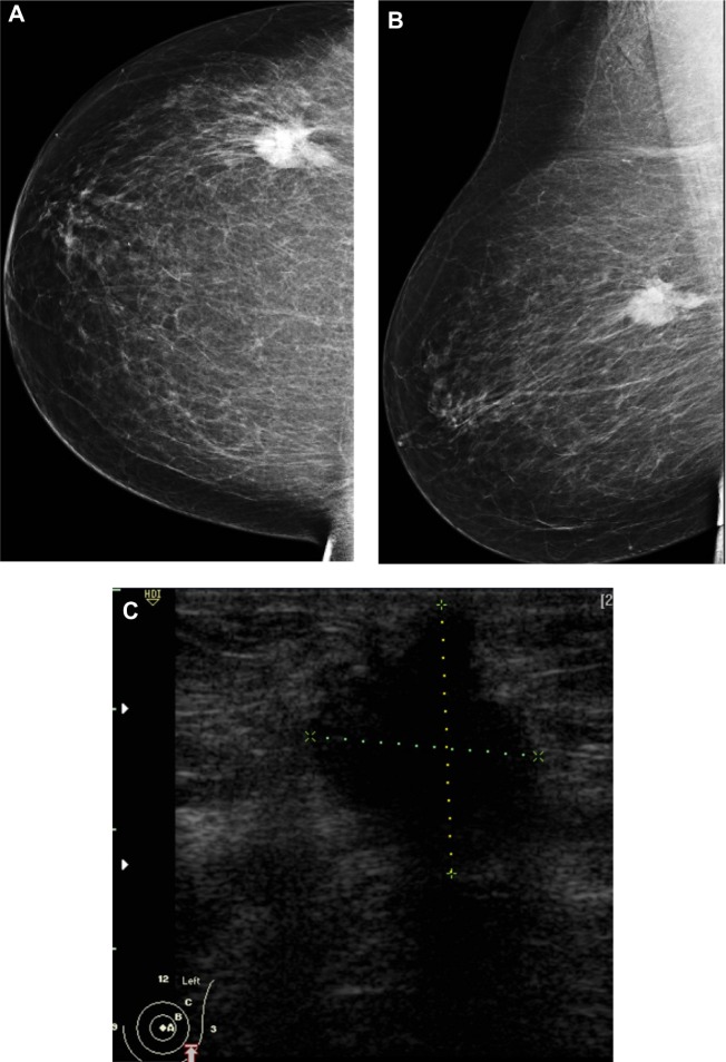
Figure 4.
A 72-year-old female with left breast mass. (A and B) Craniocaudal and mediolateral mammogram of left breast revealed an irregular spiculated, dense mass (35 × 25 mm) in the upper outer quadrant close to the pectoral muscle. Single benign macrocalcification is noted in the central area. (C) Ultrasound of the left breast revealed a spiculated, hypoechoic, solid mass (30 × 23 mm) with strong acoustic shadow at 3–4 o’clock.
Note: The mass extends to the pectoralis muscle with its long axis perpendicular to the skin.
Abbreviations: CC, craniocaudal; ML, mediolateral.

