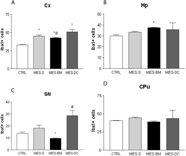
Figure 3.
Quantification of microglia expressing Iba1 in four brain structures of naïve medium- and bone marrow-injected CTRL, MES-S, MES-BM and MES-DC animals: (A) cortex (Cx); (B) hippocampus (Hp); (C) substantia nigra (SN); and (D) striatum (CPu). A significant reduction in the number of Iba1 cells was detected in the Cx (A), Hp (B) and SN (C) for f the MES-BM group (In A *p < 0.05 versus CTRL group; #p < 0.05 versus MES-DC group; in B *p < 0.05 versus CTRL and MES-DC groups; in C *p < 0.05 versus the three groups; #p < 0.05 versus CTRL). ANOVA one-way followed by Tukey’s multiple comparison test and Kruskal-Wallis, followed by Dunn’s test. Data are presented as the mean ± SEM.

