
Figure 5. The predicted three-dimensional models of U. unicinctus SDO based on SDO protein in A. thaliana (PDB ID: 2GCU).
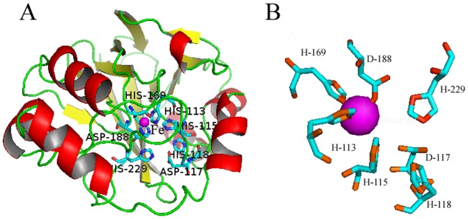
A. The β-strands, α-helices, and loop regions are shown as yellow, red, and green ribbons, respectively. The typical β-lactamase fold and metal binding sites are labeled. B. The iron (magenta sphere) binding amino acids, H113, H169 and D188, in metal binding site I form the 2His:1Asp facial triad, the remaining residues shown are found in the metal binding site II around the iron.