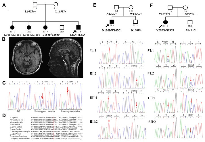
Figure 1. The pedigrees, brain MRIs, and CHIP mutations identified.
(A) The pedigree of family 1 with autosomal-recessive spinocerebellar ataxia. (B) The brain MRI of II-5 in family 1. Panel (left): axial T1-weighted image showing atrophy of the cerebellar vermis. Panel (right): midline sagittal T1-weighted image showing cerebellar atrophy, particularly evident in the superior vermis, with enlargement of the fourth ventricle. (C) Sanger sequencing results of codons 164–166 in exon 1 of the CHIP gene in a WT subject (left), an individual carrying the heterozygous variant (middle), and an individual carrying the homozygous c.493C>T (p.L165F) mutation (right). (D) The L165F missense mutation occurred at an evolutionarily conserved amino acid (in red) in the CHIP. (E and F) The pedigrees of families 2 and 3. Sanger sequencing results of the members of these two families. The red arrows indicate the mutation sites.

