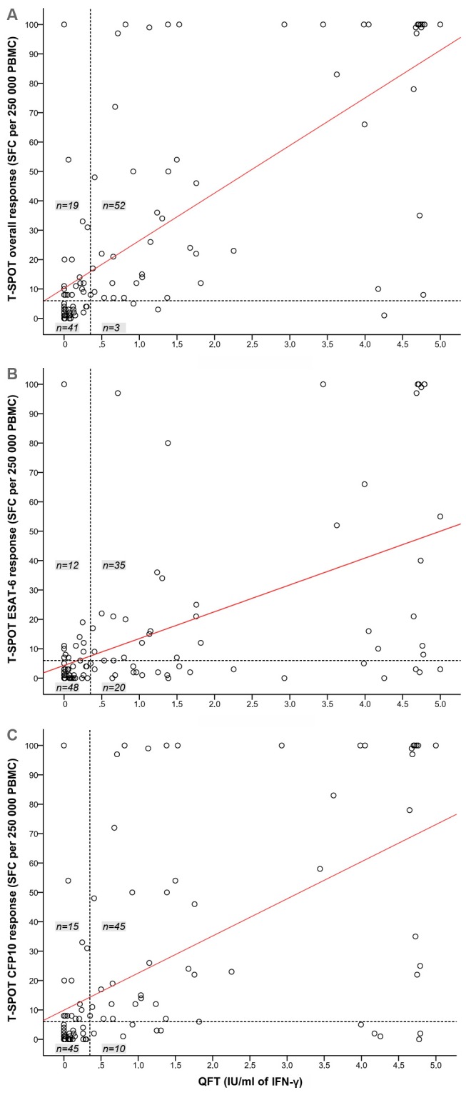
Figure 2. Distribution of quantitative T-SPOT and QFT responses.

(A) T-SPOT overall, (B) T-SPOT ESAT-6, and (C) T-SPOT CFP10 responses plotted against the quantitative QFT IFN-γ response. T-SPOT responses >100 SFC are shown as 100 SFC. QFT IFN-γ responses >5.0 IU/ml are shown as 5.0 IU/ml. The dashed horizontal and vertical lines represent the diagnostic cut-offs of 0.35 IU/ml (QFT) and 6 SFC (T-SPOT). The red diagonal line represents the regression line.
IFN-γ: interferon-γ; PBMC: peripheral blood mononuclear cells; QFT: QuantiFERON®-TB Gold In-Tube; SFC: spot forming cells; T-SPOT: T-SPOT®.TB.
