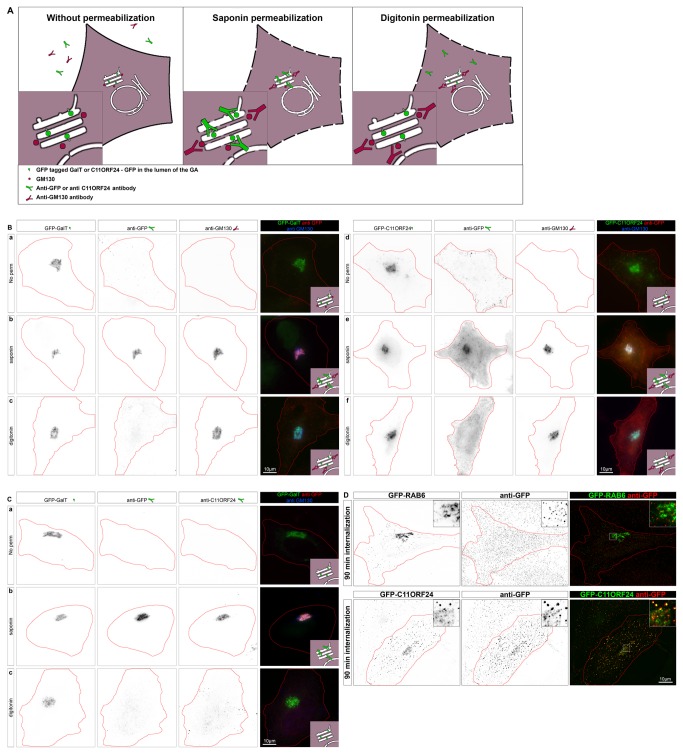
Figure 3. C11ORF24 has a long luminal domain and a short cytosolic tail.
(A) Cartoon of a cell expressing GFP-tagged GalT or C11ORF24 and stained with an anti-GFP antibody and an anti-GM130 antibody. When the cells are not permeabilized (left) the antibodies don’t bind their targets. After saponin permeabilization (middle) all antibodies are able to reach their targets since all membranes are permeabilized. After digitonin permeabilization (right) only the cytosolic epitopes (GM130) are labeled whereas the luminal epitopes (GFP of GalT or C11ORF24) are protected by the intact Golgi membranes. Protocol modified from [18] (B) HeLa cells expressing GFP-GalT (a-c) or GFP-C11ORF24 (d-f) were stained with GFP and GM130 antibodies prior to fixation (a, d) or after fixation and permeabilization with saponin (b,e) or after fixation and permeabilization with digitonin (c,f). The GFP of C11ORF24 is accessible for the anti GFP antibody only after permeabilization of the Golgi membrane. (C) HeLa cells expressing GFP-GalT were stained with GFP and C11ORF24 antibodies prior to fixation (a) or after fixation and permeabilization with saponin (b) or after fixation and permeabilization with digitonin (c). The epitope of the C11ORF24 antibody is accessible only after permeabilization of the Golgi membrane. (D) HeLa cells were either transfected with GFP-RAB6 (line 1) or GFP-C11ORF24 (line 2) 18h prior to the incubation with the anti-GFP and internalization was performed at 37°C for 90 minutes. Cells were then fixed and stained with a secondary antibody and the localization of the anti-GFP antibody was compared with the GFP signal.

