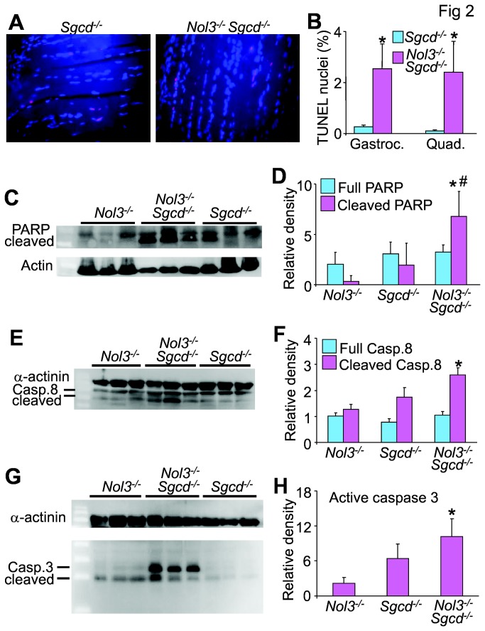
Figure 2. Arc deficiency in Sgcd-/-Nol3-/- mice alters some markers of muscle apoptosis.
A, Images of TUNEL staining (red) in longitudinal sections from quadriceps of 4 week-old mice. DAPI shows nuclei in blue. B, Quantification of TUNEL positive nuclei in gastrocnemius and quadriceps, which was determined by taking the relative proportion of TUNEL positive to normal nuclei only in a skeletal muscle fibers. *P<0.05 vs Sgcd-/-; N=6 per group with the identical 4 quadrants of the muscle counted per group. C, Western blot and D, quantification for the 85 kDa fragment of cleaved PARP from quadriceps lysates of Nol3-/-, Sgcd-/- and Nol3-/-Sgcd-/- mice (Actin serves as a loading control). *P<0.05 vs Nol3-/-; #P<0.05 vs Sgcd-/-; N=3 per group. E, Western blot and F, quantification of full-length (57 kDa) and cleaved caspase 8 (43 kDa) from quadriceps lysates of Nol3-/-, Sgcd-/-, and Nol3-/-Sgcd-/- mice (α-actinin serves as a loading control). *P<0.05 vs Nol3-/-; N=3 per group. G, Western blot and H, quantification of cleaved caspase 3 (17 and 12 kDa) from quadriceps lysates of Nol3-/-, Sgcd-/-, and Nol3-/-Sgcd-/- mice (α-actinin serves as a loading control). *P<0.05 vs Nol3-/-; N=3 per group.

