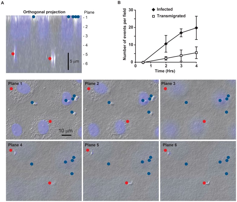
Figure 1. T. cruzi migrates across an endothelial monolayer.
(A) Endothelial cells (EC) grown on three dimensional collagen matrices were fixed and stained with DAPI to visualize nuclei. Samples were imaged using phase contrast and immunofluorescence microscopy. A typical control sample is shown. Parasites that had infected the ECs (blue dot) were found in the same focal plane (Plane 1 in the image) as EC nuclei (shown in blue). Parasites that underwent transendothelial migration were identified based on their morphology within the collagen matrix up to ∼10 µm below the EC monolayer (red dot, Planes 4 and 6). A projection of the cross-section is shown for orientation. Typically, H&E was used to score, DAPI stained sections with phase are shown here for clarity. (B) To monitor the time course of infection and transmigration separate plates of control samples were incubated for the indicated times before the reaction was stopped by fixation and quantified as described in the materials and methods. Data shown were collected from three experiments with multiple replicates per experiment.

