
Fig. 4.
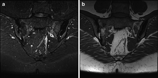
Coronal images of the SIJs demonstrating typical findings of acute or chronic sacroiliitis with sclerosis, fatty and oedema-like change as shown on STIR (a) and T1w (b) images. The diagnosis is not in question
Official websites use .gov
A
.gov website belongs to an official
government organization in the United States.
Secure .gov websites use HTTPS
A lock (
) or https:// means you've safely
connected to the .gov website. Share sensitive
information only on official, secure websites.
Coronal images of the SIJs demonstrating typical findings of acute or chronic sacroiliitis with sclerosis, fatty and oedema-like change as shown on STIR (a) and T1w (b) images. The diagnosis is not in question