
Fig. 1.
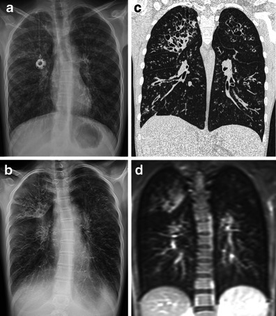
A 15-year-old girl with cystic fibrosis examined with the four different imaging modalities within 4 months. A frontal radiograph (a) acquired on the same occasion as the tomosynthesis examination (b). CT (c) performed four months earlier and MRI (d) performed 2 weeks earlier (T1-weighted sequence, with intravenous contrast). Tomosynthesis, CT and MRI sections were selected to represent approximately the same imaging plane, to simplify the comparison between the modalities. Overinflation, bronchial wall thickening, bronchiectasis and mucus plugging were present in both lungs, with the most severe changes in the right upper lobe