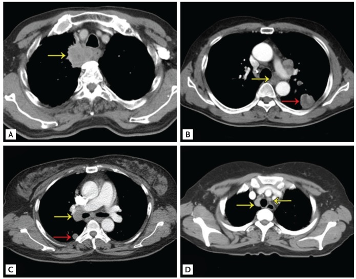
Figure 1.
(A) A chest computed tomography (CT) scan reveals a large paratracheal mass (yellow arrow). Transbronchial needle aspiration was recommended instead of percutaneous core needle biopsy (PCNB). (B) Simultaneous diagnosis and staging of a suspected primary lesion (red arrow) by CT scan. PCNB was recommended due to accessibility issues. The CT scan also revealed a lesion suspicious for mediastinal lymph node (LN) metastasis (yellow arrow). (C) CT scan reveals a small-sized nodule in the right lower lobe (RLL) of the lung (red arrow) and LN enlargement in the right Hilar area (yellow arrow). The RLL nodule is too small for PCNB. (D) CT scan reveals LN enlargement only at the anterior/posterior window (yellow arrows), and so mediastinoscopy is recommended.

