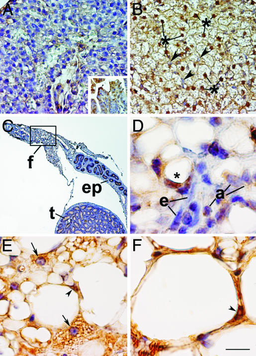
Fig. 5.
Differential expression of pRB during development of WAT and BAT and during transdifferentiation of white into brown adipocytes. (A) BAT anlage of an embryonic day 19 mouse fetus. No nuclear pRB immunoreactivity is observed. (Inset) Internal positive control of the same fetus showing pRB-positive nuclei of apical cells of intestinal villi (arrowheads). (B) BAT of a 10-day-old mouse. Nuclei of well differentiated brown adipocytes are pRB-positive (*), and nuclei of endothelial cells are negative (arrowheads). (C) Epididymal fat pad of a 9-day-old mouse. f, fat pad; ep, epididymus; t, testis. (D) Epididymal WAT of a 9-day-old mouse (enlargement of the squared area in C). Adipocyte precursors with lipids droplets (*) show nuclear staining. Endothelial cells (e) and adipoblasts (a) are pRB-negative. (E) Retroperitoneal WAT of a 20-week-old rat treated with the β3-adrenergic agonist CL-316243 for 7 days. Most of the transdifferentiating multilocular adipocytes exhibit pRB negative nuclei (arrows). A unilocular adipocyte with positive nucleus is visible (arrowhead). (F) Same tissue as in E. A unilocular adipocyte with positive nucleus is visible (arrowhead). (Bars: A and B = 30 mM; A Inset = 60 mM; D and F = 10 mM; C = 200 mM; E = 15 mM.)

