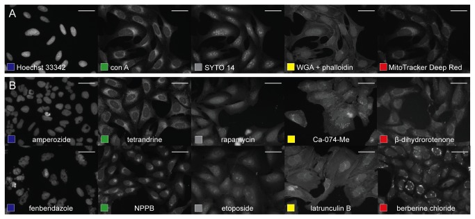
Figure 1. The cell-painting assay applied to U2OS cells.
(A) Cells labeled with Hoechst 33342 (nuclei, blue), concanavalin A (ER), SYTO 14 (nucleoli), phalloidin (actin), WGA (Golgi), MitoTracker Deep Red (mitochondria). Scale bars 50 µm. (B) Ten diverse phenotypes in compound-treated U2OS cells: toroid nuclei (amperozide); giant, multinucleated cells (fenbendazole); abundant ER (tetrandrine); redistribution of ER to one side of nucleus (NPPB); reduced nucleolar size (rapamycin); large, flat nucleoli (etoposide); bright, abundant Golgi staining (Ca-074-Me); actin breaks (latrunculin B); extensive mitochondrial fission (Beta-dihydrorotenone); and redistribution of mitochondria (berberine chloride). Scale bars 50 μm.

