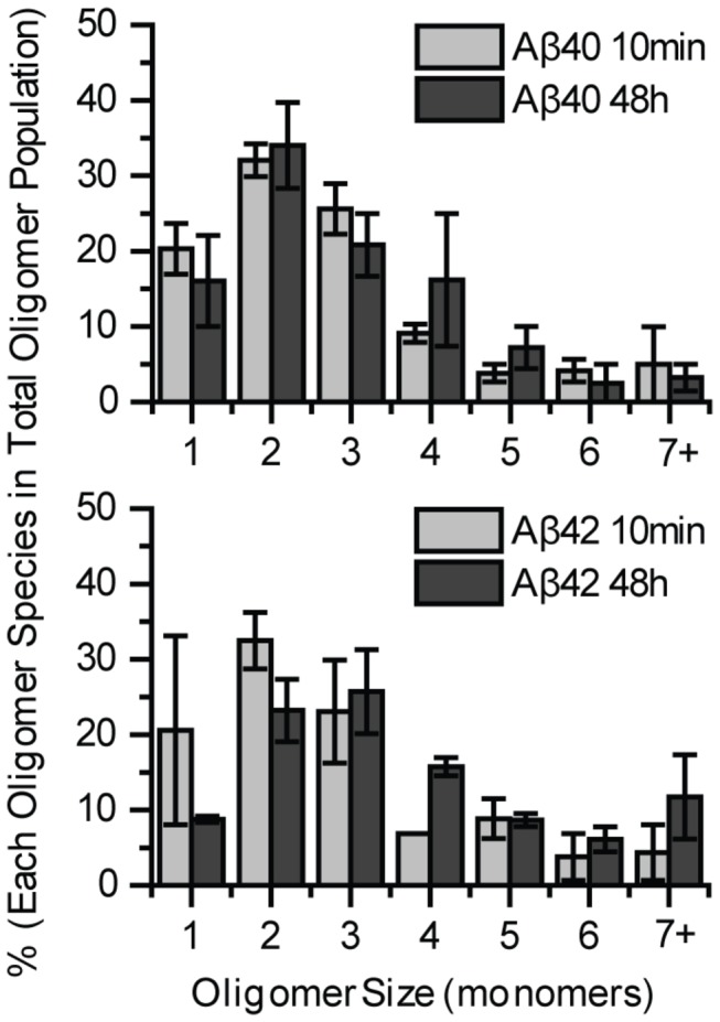
Figure 2. Aβ40 or Aβ42 oligomers form mainly dimers and show little growth on neurites.

2nM Aβ40-HL555 or Aβ42-HL647 was incubated with primary hippocampal neurons for 10 minutes and 48 hours before imaging. Comparison of the oligomeric size distribution between 10 minutes and 48 hours shows limited growth for both Aβ40-HL555 (Mann-Whitney U test, p > 0.1) and Aβ42-HL647 (Mann-Whitney U test, p = 0.001). The distribution is normalized to total Aβ oligomers. Percentages of each condition were calculated from two different experiments, 5 images each. Each image contained at least 50 oligomers. Error bars represent standard deviation of the mean. The percent is obtained by normalizing to the total number of oligomers.
