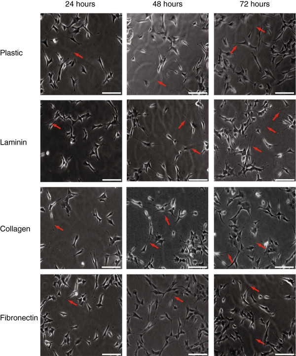
Figure 1.
Optimisation of matrices for differentiation of SH-SY5Y cells. SH-SY5Y cells were plated on 6 well plates uncoated (plastic) or coated with 10 μg/ml of laminin, collagen or fibronectin. Cells were incubated in regular DMEM media containing 10% FBS for 24, 48 or 72 hours. Pictures were taken at 20× using Metamorph software. Scale bar = 20 μm.

