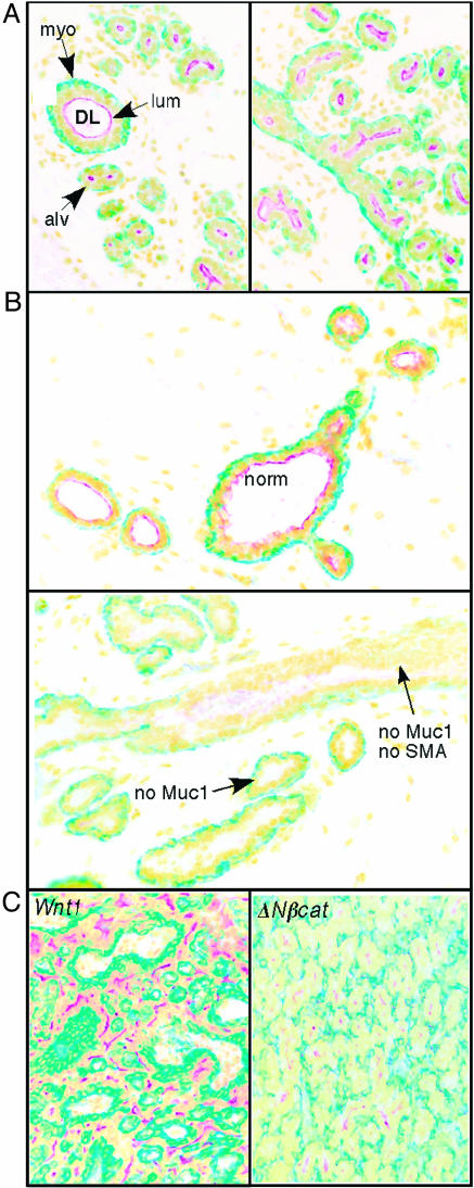
Fig. 1.
Wnt1- and ΔNβcat-induced tumors contain at least three types of cells, including cells that express markers of luminal or myoepithelial cells and some that express neither. Samples of normal mammary gland from a 12-day pregnant mouse (A), hyperplastic mammary gland from an 11-week-old virgin Wnt1-transgenic mouse (in estrus) (B), and Wnt1- or ΔNβcat-induced mammary tumor (C) were fixed and paraffin-embedded, and the sections were stained with antibodies to Muc-1 (stains the apical membrane of luminal cells; red) or to SMA (stains the cytoskeleton of myoepithelial cells; green). Nuclei were counterstained with Hoecsht (shown here in yellow). (A) Mature mammary glands comprised of hollow ducts (ductal lumen; DL) lined by an inner luminal layer (lum) and a basal myoepithelial cell layer (myo). During growth and differentiation, alveoli (alv) develop that have smaller lumens and an incomplete basal layer. (B) Hyperplastic glands are characterized by core ductal zones that have relatively normal architecture (norm; Upper), surrounded by pseudolobuloalveolar elements with multilayered epithelia that sometimes entirely fill the ductal lumen. There is no accumulation of apical Muc-1 in these zones of hyperplasia, and the basal layer expresses SMA only in patches. (C) One representative example each of a Wnt1- and a ΔNβcat-induced tumor are shown. Cells are organized into cohesive sheets of myoepithelia interspersed with bands of luminal derivatives. This pattern is typical of the majority of tumors; the remaining 15% of tumors were undifferentiated, in papillary form, and infiltrated with highly reactive stroma.

