Fig. 2.
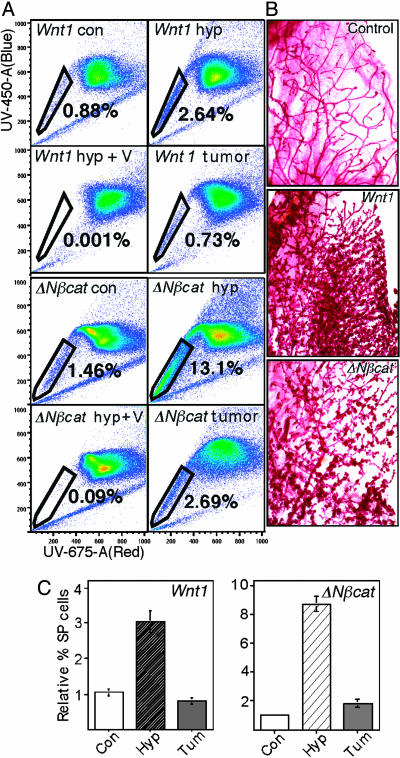
Progenitor cell-enriched SP fractions are increased in Wnt1- and ΔNβcat-transgenic mammary glands. MECs were prepared from mammary glands from 2- to 3-month-old FVB MMTV-Wnt1- and MMTV-ΔNβcat-transgenic mice, together with control littermates (this experiment shows pooled MECs from 7 Wnt1:FVB compared with 11 control FVB mice and 11 ΔNβcat:FVB compared with 12 FVB controls). (A) Cells were labeled with Hoecsht dye and assayed by flow cytometry for fluorescence in the red and blue channels. The side population was gated as a spur of cells with less fluorescence. Representative scans are shown for two sets of samples. The first shows control Wnt1+/– hyperplastic glands (Wnt1 hyp), the hyperplastic cells labeled in the presence of verapamil (an inhibitor of Hoechst dye efflux), and a Wnt1-induced tumor. The second set shows the same sample set for ΔNβcat-induced glands and a tumor. (B) The morphogenesis of control, Wnt1-transgenic glands and a [ΔNβcat]-transgenic gland (3 months old) is shown in carmine-stained whole-mount preparations. (C) The results of SP assays were quantified. Average values for the relative proportion of SP cells, together with their SDs, were derived from three independent sorts, and these results were repeated with a separate set of MECs isolated on different days. Analysis of SP fractions of [MMTV-Wnt]:C57BL6 transgenic mammary glands gave similar results. Con, control; hyp, hyperplastic; tum, tumor.

