
Figure 5.
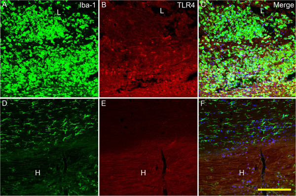
Immunofluorescent labeling for TLR4 and IBa-1 in the lesion site and the hematoma at 3 days post injury. In the lesion site (A-C), there were abundant IBa-1 positive cells, quite part of which express TLR4, showed by confocal microscopy. Most of the microglia/macrophages were round with short or blunt processes. To the contrast, in the distal hematoma and the area adjacent to it (D-F), there were few TLR4 positive cells, and IBa-1 labeled cells were small with thin processes. TLR4 immunoreactivity: red; IBa-1 immunoreactivity: green; Hoechest 33342: blue. Bar = 200 μm.