Figure 3.
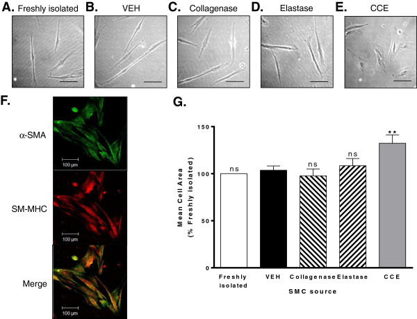
PCA SMC morphology. Cells were explanted from both freshly isolated PCA and bioreactor vessels and maintained in cell culture in full growth medium. Representative phase contrast images of cells explanted from (A) freshly isolated tissue (B), VEH, (C) collagenase, (D) elastase and (E) CCE-pre-treated vessels, scale bar = 100 μm. (F) Immunocytochemical staining for α-SMA (green) and SM-MHC (red) and co-localisation (orange). Magnification × 400, scale bar = 100 μm. (G) Mean cell areas of 100 individual cells per condition were quantified using Image J and expressed relative to the matched freshly isolated vessel. All n = 3, ns = non significant, **P < 0.01.

