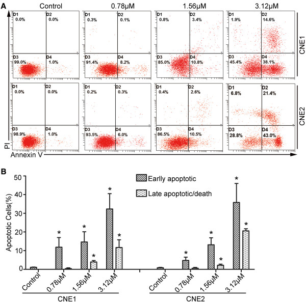
Figure 2.
LK-A induces apoptosis of human nasopharyngeal carcinoma cells. Flow cytometry analysis of CNE1 and CNE2 cells treated with 0.78, 1.56 and 3.12 μΜ LK-A for 48 hrs. (A) Dot plots showing the percentage of viable (D3), early apoptotic (D4), late apoptotic (D2) and necrotic (D1) cells. (B) Bar chart indicating the increased proportion of early and late apoptotic cells after treatment with LK-A. Data are shown as the mean ± SD from two independent experiments. *p < 0.05 vs. control group (untreated).

