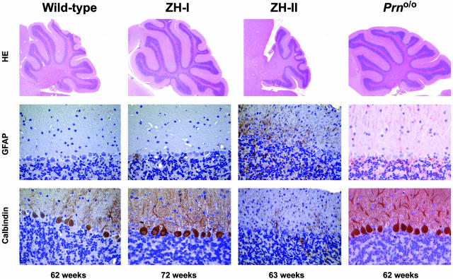
Fig. 5.
Cerebellar histopathology of Prnp and Prnd mutant mice. (Top) Parasagittal sections of wild-type, ZH-I, ZH-II, and Prno/o mice stained with hematoxylin/eosin. (Middle) Glial fibrillary acidic protein (GFAP) staining of cerebellar cortex with gliosis in ZH-II mice, but not in wild-type, ZH-I, or Prno/o mice. (Bottom) Calbindin stains (specific for PC). Wild-type, ZH-I, and Prno/o mice show an intact PC layer, whereas ZH-II mice undergo extensive cell loss.

