Fig. 1.
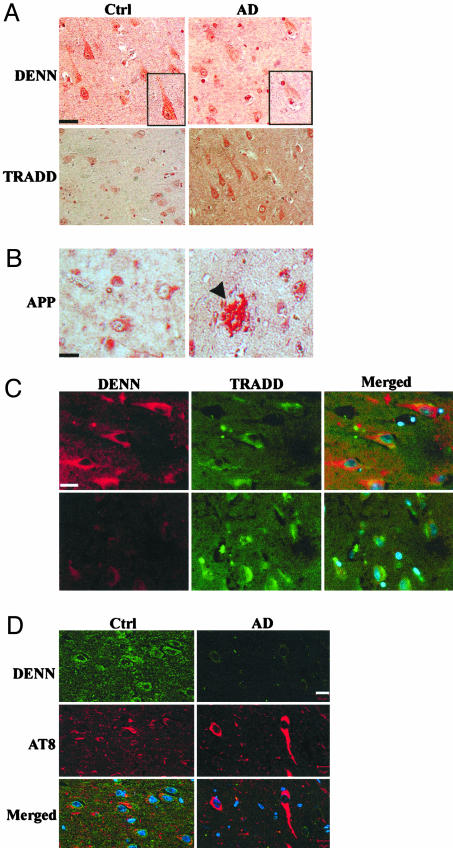
DENN expression is reduced in AD-affected human hippocampal neurons. (A) CA1 neurons from human hippocampus. Immunoperoxidase staining of DENN in control and AD hippocampus reveals cytoplasmic and membrane staining and faint cytoplasmic and nuclear staining, respectively. TRADD expression is increased in AD relative to the control. (Bar, 20 μm.) (B) Affected CA1 region of hippocampus shows Aβ plaque formation (arrowhead) and increased APP immunostaining. (C) Immunocytochemical colocalization of DENN and TRADD. DENN is stained with Alexa594 (red), and TRADD is stained with Alexa 488 (green). DENN is decreased and TRADD increased in AD relative to control. DENN and TRADD show minimal, focal overlap in the perinuclear cytoplasm of pyramidal neurons in controls but not in AD. (Bar, 20 μm.) (D) CA1 region of human hippocampus was stained with antibodies specific to DENN (Alexa 488, green) and hyperphosphorylated tau, mAb AT8 (Alexa 594, red). Normal, aged control shows DENN is localized to the cytoplasm of pyramidal neurons and surrounding neuropil. DENN expression is diminished in AD in merged, confocal images, compared to control neurons. Nuclei are labeled with Hoechst 33342 (blue). (Bar, 10 μm.) Filter settings were equivalent in both control and AD tissues.

