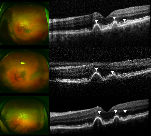
Figure 3.

Patient 2. (Top) Day 0, multiple nodular hyperreflective signals in the outer retina are indicated by the arrowheads. The rightmost arrowhead points to hyperreflective signals at PRL and RPE level. RPE disruption is present due to drusen. There are sheets of vitreous cells shown in the Optos® wide-angle image. (Center) Day 77, arrowheads indicate hyperreflective changes at PRL and RPE. Severe vitritis as shown in the left fundus contributes to the noisy SD-OCT scan. Optos® wide-angle image shows increased vitreous inflammation. (Bottom) Day 119, arrowheads point to nodular hyperreflective signals that may be lymphomatous infiltrates. The view of the fundus is clearer post-vitrectomy.
