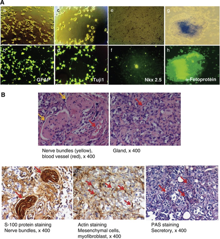
Figure 2.
Pluripotency of bovine iPSCs. (A) In vitro differentiation of and marker expression by bovine iPSC-derived ectodermal, mesodermal, and endodermal precursor cells. Immunostaining with antibodies directed against the astrocyte-specific antigen GFAP (ectodermal differentiation), neuron-specific antigen Tuj1 (ectodermal differentiation), cardiomyocyte-specific antigen Nkx 2.5 (mesodermal differentiation), or α-fetoprotein (endodermal differentiation). (B) Teratoma formation 6–8 weeks after the transplantation of bovine iPSCs into SCID mice. Teratomas were sectioned and stained with hematoxylin and eosin. Immunohistochemical staining was performed using antibodies specific for S-100 (nerve bundles) and muscle-specific actin (mesenchymal cells and myofibroblasts) or PAS staining (secretory cells) ( × 400 magnification). In panel a, the red and yellow arrows indicate blood vessels and nerve bundles, respectively. In panel b, the red arrows indicate glands. S-100 staining indicates nerve bundles (panel c; red arrows), and muscle-specific actin staining indicates mesenchymal cells and myofibroblasts (panel d; red arrows). PAS staining indicates secretory cells (panel e; red arrows). The proliferation index of the whole teratoma was<3%

