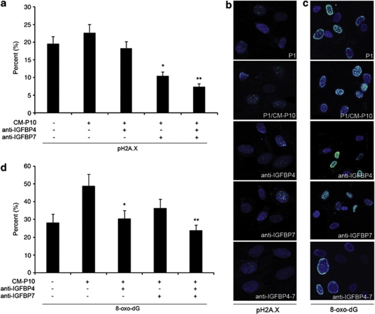
Figure 4.
Reversal of the DNA-damage response of young MSC induced by CM-P10 by IGFBP4 and IGFBP7 blocking. (a and b) Phosphorylated H2A.X staining performed on young MSC cultured with untreated and IGFBP4 and/or IGFBP7 antibody-treated CM-P10; *P<0.05; **P<0.01 versus P1 MSC grown with untreated CM-P10. Fluorescence micrographs showing merge of representative fields of cells stained with anti-pH2A.X (green) and Hoechst 33342 (blue). (c and d) 8-oxo-dG staining performed on young MSC cultured with untreated and antibody-treated CM-P10. *P<0.05, **P<0.01 versus P1 MSC grown with untreated CM-P10. Fluorescence micrographs showing merge of representative fields of cells stained with anti-8-oxo-dG (green). Nuclei were counterstained with Hoechst 33342 (blue)

