Figure 1.
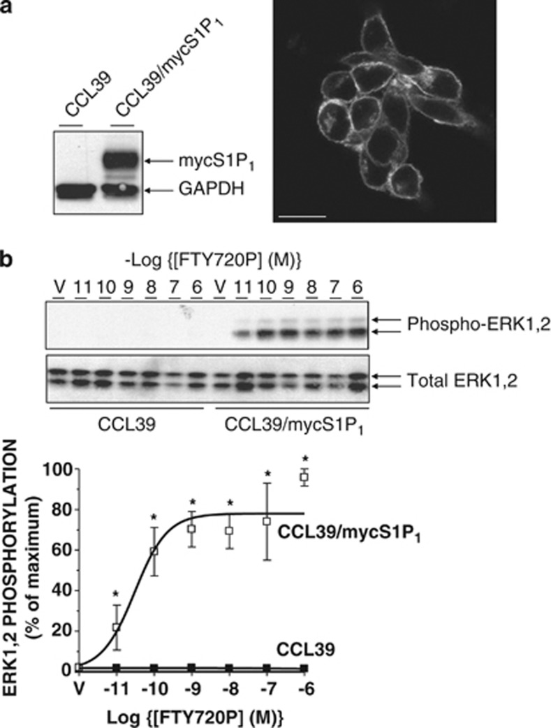
Functional expression of human S1P1 in CCL39 cells. (a) Left: detergent-soluble cell extracts from control and S1P1-expressing CCL39 cells were equalised for protein content before immunoblotting with anti-myc antibody 9E10 (to detect myc epitope-tagged receptor) and GAPDH. Right: S1P1-expressing CCL39 cells were fixed and permeabilised for staining with 9E10 antibody and Alexa 488-conjugated goat anti-mouse IgG before visualisation by confocal microscopy. Scale bar=20 microns. (b) Upper: control and S1P1-expressing CCL39 cells were treated for 5 min with the indicated concentrations of S1P receptor agonist FTY720P or vehicle (V) before the preparation of detergent-soluble cell extracts. Samples were equalised for protein content before fractionation via SDS-PAGE and subsequent immunoblotting with anti-Thr202/Tyr204 phospho-specific ERK1,2 antibodies as a surrogate marker of intracellular signalling. Equal protein loading was assessed by determining total ERK1,2 levels. Lower: data are presented as mean values±S.E. for n=3 separate experiments. *P<0.05 versus identically treated CCL39 controls

