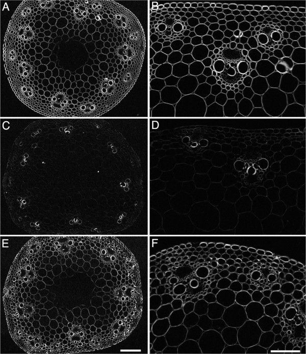
Figure 9.
Polarized-light micrographs of stem internode transverse cross-sections. Representative images of (A, B) wild type; (C, D)amiR-CESA4; (E, F)amiR-CESA7. Left hand panels are observed through a 4x lens and a gray scale value of 255 indicates a retardance value of 5 nm; Right hand panels are observed through a 20x lens and a gray scale value of 255 indicates a retardance value of 13 nm. Bars = 50 μm (E) and 100 μm (F).

