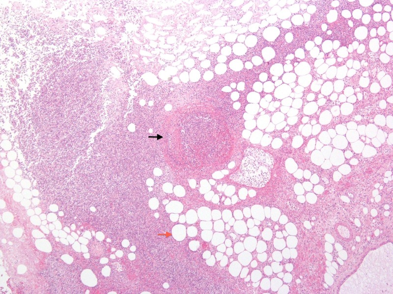
Figure 3.

Photomicrograph of necrotic perianal tissue from a subcutaneous layer biopsy. Surrounding neutrophils can be seen around non-viable adipocytes (red arrow). A thrombosed central vessel is present within the necrotic soft tissue (black arrow).
