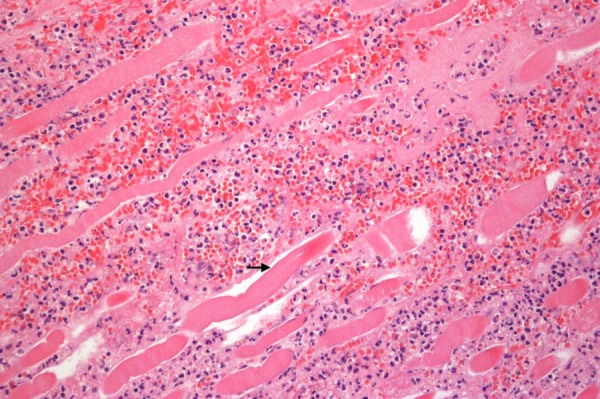
Figure 4.

Photomicrograph from the initial wound debridement demonstrating necrotising fasciitis of the smooth muscle layer. Non-viable myocytes (black arrow) are seen with broad bands of polymorphonuclear cell infiltrate along the major fascial planes.
