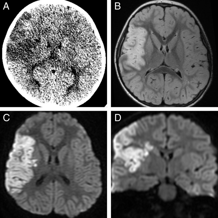
Figure 1.
Initial diagnostic imaging demonstrates right middle cerebral artery confluent low density on non-contrast head CT (A) that is confirmed to be infarction on MRI with T2 prolongation on T2 FLAIR (B) and reduced diffusion in both the axial and coronal diffusion-weighted images (C and D). There was no evidence of hemorrhage on gradient recalled echo imaging (not shown).

