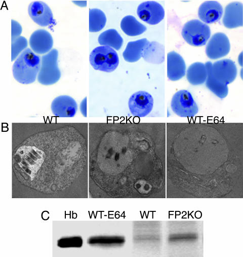
Fig. 4.
Evaluation of trophozoite hemoglobin hydrolysis. Early (24-h) trophozoites of FP2KO, WT, and E-64-treated (10 μM, beginning at 0 h) (WT-E64) parasites were evaluated by Giemsa staining (A) and electron microscopy (B). Erythrocytes infected with equal numbers of these trophozoites were lysed with saponin, washed to remove erythrocyte hemoglobin, and solubilized in reducing sample buffer, and proteins were resolved by SDS/PAGE and stained with Coomassie blue (C). Hb represents a hemoglobin control.

